Having an x-ray of your mouth is often a vital part of your visit to the dentist. But because x-rays involve exposure to radiation, some patients wonder, “Are dental x-rays safe?” Many ask:.
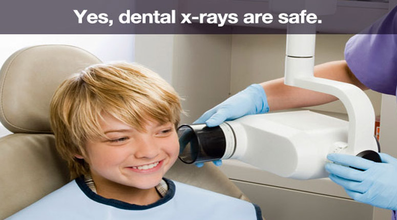
Dental x-rays are one of the safest and lowest radiation dose treatments performed. A routine exam, which includes four bitewings, is about 0.005 mSv. That’s about the same amount of radiation you’d get on an average day from the. A panoramic dental x-ray, which goes around your entire head, has approximately twice that amount of radiation.
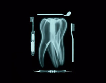
X-rays allow your dentist to see bones, tissue, and hidden surfaces of your teeth that can’t be seen with the naked eye during a visual exam.
Dental x-rays provide invaluable information to a dentist about your oral health such as the presence of gum disease, early-stage cavities, oral cancers and some types of tumors. If these problems remain undetected and go without treatment, they can grow into very serious dental problems that involve more extensive dentistry, time, money to fix.
The various types of x-rays used by your dentist serve specific purposes. A combination of x-rays may be necessary depending on the treatment plan outlined by your dentist. Some of the x-rays that may be suggested include:
Bite-wings are the most commonly used x-ray during an initial exam and in subsequent check-ups. These highlight the crowns of the back teeth. Dentists take one or two bite-wing x-rays on each side of the mouth. Each x-ray shows the upper and lower molars (back teeth) and bicuspids (teeth in front of the molars.)
Digital radiographs are the newest x-ray technique used by dental offices. Standard x-ray film is replaced with a flat electronic pad or sensor. The image is transferred digitally into a computer, where it can be viewed on a screen, stored, or printed out.
Even though digital x-rays produce lower levels of radiation than standard x-rays and are considered very safe, dentists still take necessary precautions to limit the patient’s exposure to radiation. These precautions include only taking x-rays that are needed, and using lead apron shields to protect the body.
Extraoral x-rays are made outside the mouth. These are considered “big picture” x-rays because they not only show the teeth, they also provide information on the jaw and skull and are often necessary for the effective treatment of TMD/TMJ.
These x-rays focus on only one or two teeth at a time. A periapical x-ray looks similar to a bite-wing x-ray, but it shows the entire length of each tooth, from crown to root.
This x-ray captures the entire mouth in a single image, including the teeth, upper and lower jaws, surrounding structures and tissues and requires a special machine. The benefit of this type of x-ray is that it eliminates the need for multiple x-rays.
If x-rays cannot be postponed until after the birth and are necessary because of a needed dental procedure, the American College of Radiology says that no single diagnostic x-ray has a radiation dose significant enough to cause adverse effects in a developing fetus.
1. Monitor your overall dental health.
2. Look at the roots of your teeth.
3. Check the health of the bone around and beneath your teeth.
4. Find cavities, even hidden ones.
5. Check on the development of erupting teeth.
Any time you have a question or concern regarding your oral health, feel free to contact Dr. Guerra at 561-844-6146, and his friendly dental team. We’re here to help!


